Zeiss Cirrus HD-OCT
Featuring spectral domain technology, Cirrus HD-OCT delivers exquisite high-definition images of ocular structures by obtaining 27000 scans per second . The retina, optic nerve, iris and cornea can be imaged with a resolution of 5 microns. All images are stored on a central server for immediate analysis of current and past exams. The retinal and optic nerve measurements are compared to a powerful control database and analyzed. Abnormal results are highlighted and given a percentage of deviation from normal. Baseline measurements are kept for future comparisons and statistical analysis.
Chronic Open Angle Glaucoma Diagnosis & Management

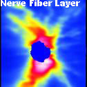
The earliest sign of glaucoma is thinning of the nerve fiber layer of the retina. The OCT is the most advanced diagnostic tool available to detect abnormalities in a patient’s nerve fiber layer. Measurements are obtained with a resolution of 5 microns and measurement reproducibility within 1.2 microns allowing better detection of new patients with glaucoma and a more objective and precise way of monitoring treatment success in patients with known glaucoma on medication. With the Cirrus HD-OCT early diagnosis of glaucoma is possible and loss of vision can be prevented.
Narrow Angle Glaucoma Detection and Prevention
A less common form of glaucoma is closed-angle, or narrow-angle, glaucoma. Closed-angle glaucoma occurs when the drainage angle of the eye becomes blocked. Unlike open-angle glaucoma, eye pressure usually goes up very fast. The pressure rises because the iris — the colored part of the eye — partially or completely blocks off the drainage angle. A closed-angle glaucoma attack is a medical emergency and must be treated immediately.
The OCT is also able to image structures in the anterior segement of the eye. Imaging the intersection of the iris and the cornea of the eye allow determination of the angle and degree of separation between the two ocular structures. Recommendations as to the benefit of Laser Iridotmy to prevent angle closure can be made with greater certainty.
Retinal Disease Detection
The macula can be scanned to diagnose and manage a variety of retinal diseases including:
- Macular Degeneration
- Macular Pucker and Epiretinal Membrane Formation
- Cystoid Macular Edema
- Macular Traction Syndrome
- Macular Hole
- Diabetic Eye Disease
- Central Serous Chorioretinopathy
- Macular Edema from Retinal Vein Occlusions
[Best_Wordpress_Gallery id=”9″]