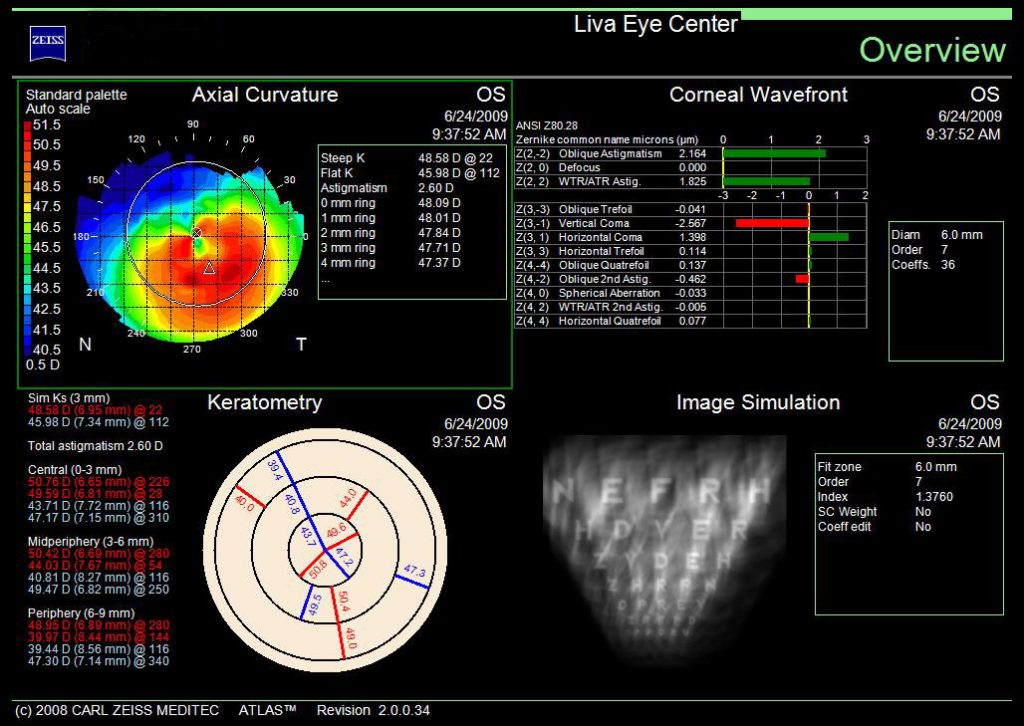
Corneal topography, also known as photokeratoscopy or videokeratography, is a non-invasive medical imaging technique for mapping the surface curvature of the cornea, the outer structure of the eye. Since the cornea is responsible for about 70% of the eye’s refractive power, its topography is of critical importance in determining the quality of vision. The procedure is carried out in seconds and is completely painless.
Placido Disk Technology is used to create a three-dimensional map that can assist in the diagnosis and treatment of a number of corneal conditions. It is useful in planning refractive surgery such as LASIK and PRK . Sophiscated sofware analysis of acquired data can indicate if corneal pathology such as Keratoconus is present which would disqualify someone for refractive surgery. It can also evaluate the cornea to aid in the fit of contact lenses. The procedure is carried out in seconds and is completely painless.
Cataract surgery patients can benefit from corneal topography for several reasons. First, it can measure the power and axis of corneal astigmatim to determine the placement of Toric IOLs which can reduce or eliminate the distortion caused by pre-existing corneal astigmatism. Secondly, it can measure higher order aberrations such as spherical aberration which reduces night vision contrast sensitivity. Customizing the selection of the proper aspheric IOLs to neutralize the spherical aberration of the cornea enhances night vision and reduces glare. Thirdly, it can detect pathological conditions which may reduce the efficacy of cataract surgery.
