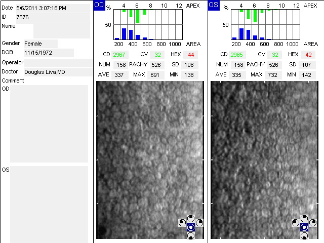
Specular microscopes are for analysis of the corneal endothelium. The endothelium is the back layer of the cornea. The endothelium pumps fluid out of the cornea enabling the cornea to be clear. Endothelial dysfunction leads to corneal edema(swelling) and reduced vision and ultimately to corneal failure requiring transplantation. Fuchs’ disease is a common hereditary corneal dystrophy that results in an abnormal endothelial layer. Specular microscopes image the abnormal corneal endothelium in Fuchs’ disease and can assess the stage of the disease.
The specular microscope at Liva Eye Center represents the latest in endothelial cell analysis, algorithms, and computer technology. Enhanced with auto-focus, auto-alignment, and auto-cell counting.
In addition, non-contact pachymetry (meaurement of corneal thickness) is performed at the same time image analysis of the corneal endothelium is aquired.
The assessment of the corneal endothelial cell layer, morphology of endothelial cells, and corneal pachymetry are all indicators of corneal health.
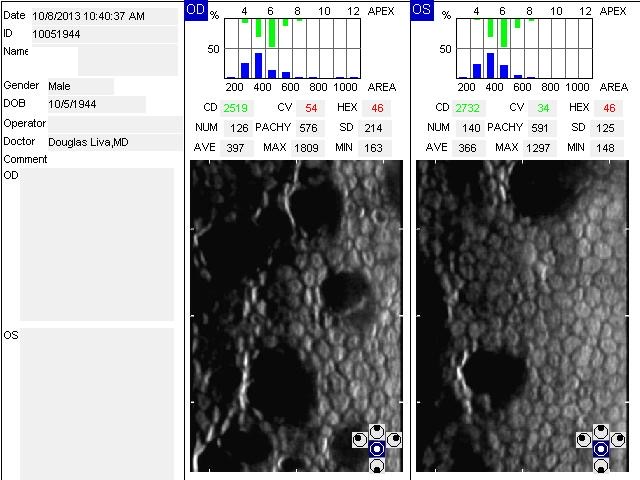
Fuchs’ Disease with Corneal Guttata