High Definition Spectral Domain Imaging
Glaucoma is one of the leading causes of vision loss in the United States. Glaucoma refers to a group of conditions in which the optic nerve has sustained damage. In most cases this is linked to an increase in intraocular pressure (IOP), although it can occur in people with normal IOP. Because there are no symptoms associated with early stage glaucoma, people often do not know they have the disease until the experience some loss of vision. For this reason, glaucoma is often referred to as a “thief of sight.”
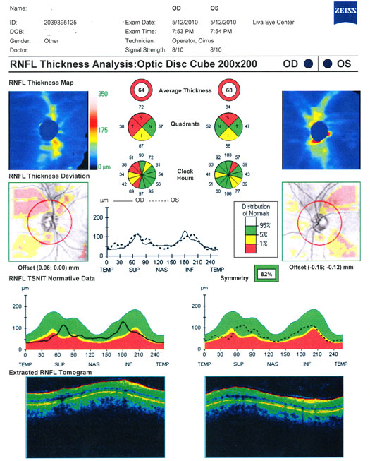
Early diagnosis of glaucoma is crucial for preventing vision loss. The earliest sign of glaucoma it the thinning of the nerve fiber layer of the retina. Dr. Liva uses the most advanced diagnostic tool available to detect abnormalities in a patient’s nerve fiber layer at the earliest possible moment. The Cirrus HD-OCT is a High Definition Spectral Domain Imaging Laser that performs 27000 scans per second of the optic nerve fiber layer. Measurements are obtained with a resolution of 5 microns. These measurements are compared to a powerful database to compare your results with normal levels for people of your gender, age, and race. The data is stored in a personal file for future reference as a baseline to allow better detection of glaucoma and also to monitor treatment success in patients with known glaucoma on medication.
With the Cirrus HD-OCT, early diagnosis of glaucoma is possible and loss of vision can be prevented.The report below illustrates a patient with a damaged nerve fiber layer due to glaucoma.
Digital Stereo Fundus Photography
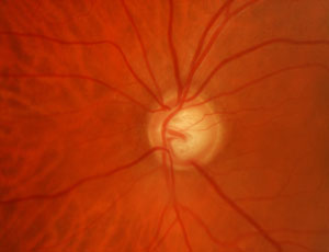
At Liva Eye Center stereophotographs of the optic nerve are taken with a high resolution digital camera to document optic disk morphology. Stereophotographs provide a detailed three dimensional image of the optic nerve which can be followed over time to detect subtle anatomical changes. Increased cupping of the optic nerve is indicative of glaucoma. In the past, detailed drawings of the optic nerve were relied upon to monitor the patient for progression of optic nerve cupping. Digital photography with computerized databases and analytical tools have rendered drawings obsolete.

Humphrey ® Field Analyzer
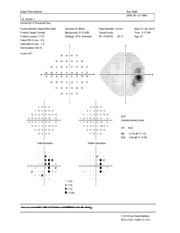
Liva Eye Center has certified ophthalmic technicians performing computerized visual field testing which is an essential component of glaucoma management. Sophisticated computer algorithms are employed to reduce testing time and improve accuracy. Normative and glaucoma databases are used to compare test results allowing statisical analysis to detect deviation from normal. The field analyzer can perform Blue-Yellow perimetry known as Short Wavelength Automated Perimetry or SWAP. SWAP has been shown to provide earlier detection of glaucomatous visual field loss than conventional white on white testing. Progression analysis provides a method to determine how effective the present level of treatment is in preventing the progression of glaucomatous visual loss. The computer software is able to quantify the degree of visual damage with a new parameter called the Visual Field index which gives an number which represents the amount of optic nerve damage a patient has sustained from glaucoma. The software can predict how much damage will be present 5 years into the future.

Selective Laser Trabeculoplasty
There are several types of glaucoma; the most common is called open-angle glaucoma. It occurs when fluid within the eye drains too slowly, causing intraocular pressure to rise. Treatment for open-angle glaucoma involves lowering IOP usually with eye drops. However, when drops fail to adequately control the eye pressure to a safe level or are not tolerated, Dr. Liva offers the Lumenis SLT laser to perform an advanced procedure. SLT stands for Selective Laser Trabeculoplasty. The “Selective” in SLT is very important. It means that the laser is used to target only specific cells (those that contain pigment) while leaving other cells unharmed. These pigment cells are found within the “clogged” Trabecular Meshwork which is the area of fluid drainage from the eye. The targetting of these cells results in increased drainage of fluid from the eye resulting in a lower eye pressure. The SLT procedure is an effective alternative to medication or more invasive surgery.
Yag Laser Iridotomy
Dr. Liva performs Yag Laser peripheral iridotomy for patients at risk for developing an attack of angle closure glaucoma due abnormality of the anatomy of the anterior segment of the eye, known as narrow angle glaucoma. Narrow angle glaucoma is a condition where the peripheral part of the iris is located in a position that can lead to the sudden blockage of fluid out of the eye causing an extreme elevation in pressure which if left untreated causes irreversible vision loss. Dr. Liva performs gonioscopy and anterior segment imaging to determine if there is an abnormality of a patient’s iris anatomy that could predispose the patient to the development of narrow angle glaucoma. The abnormaltity can be effectively treated fwith one laser session for each eye.
Please schedule an appointment with Dr. Liva as soon as possible. Whether or not you have been diagnosed with glaucoma already, he can help you take the necessary steps to preserve your vision.
